


Tomosynthesis vs Mammogram: What's the Difference?
In the ever-evolving landscape of medical technology, the detection and diagnosis of breast cancer have seen remarkable advancements, notably through the development of mammography and tomosynthesis.
These two breast imaging techniques stand at the forefront of early cancer detection, each with unique capabilities and benefits.
Mammography, a tried and tested method, has been instrumental in saving countless lives through early detection. On the other hand, tomosynthesis represents a significant leap forward, offering enhanced image clarity and accuracy, especially in detecting cancer within dense breast tissue.
Join us as we delve into tomosynthesis vs mammography, analysing their effectiveness, detection rates and image detail.
What is Mammography?
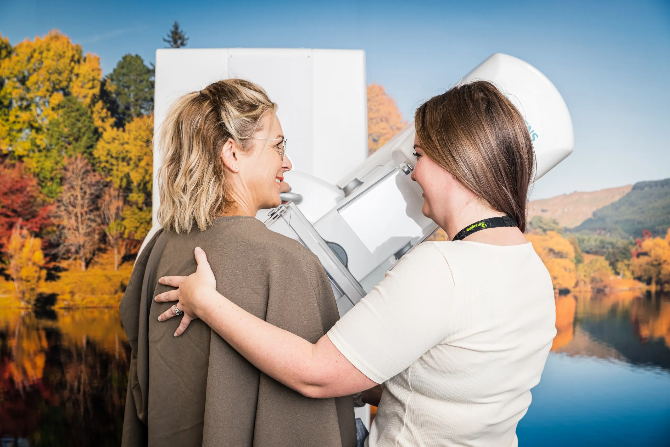
Mammography, a specialised medical imaging technique, utilises low-dose X-rays to examine the human breast, serving as a critical tool in the early detection and diagnosis of breast cancer.
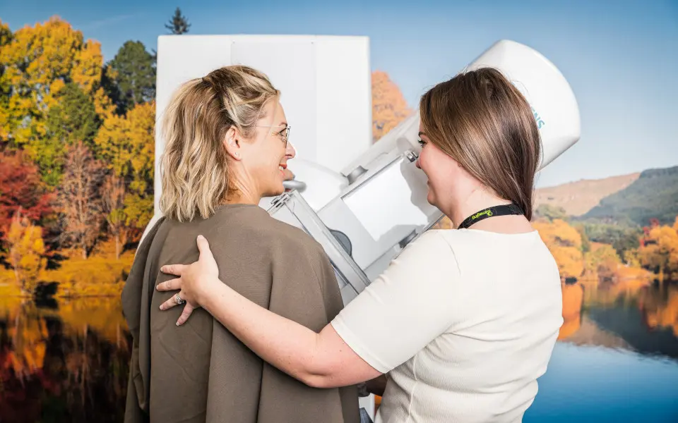
Mammography originated in the early 20th century, with significant advancements over the decades enhancing its efficacy and safety. During a mammogram, the breast is gently compressed between two plates to spread the breast tissue, ensuring the X-ray images are as clear as possible. A radiologist then examines these images for any signs of abnormality, such as lumps or microcalcifications, that could indicate the presence of breast cancer.
The role of mammography in breast cancer screening cannot be overstated; it has been instrumental in reducing mortality rates by enabling the early detection of cancer, often before symptoms appear. Through routine screening mammograms, it's possible to identify breast cancer at an early stage, when treatment is more likely to be successful, underscoring the vital role of mammography in women's health initiatives worldwide.
What is Tomosynthesis?
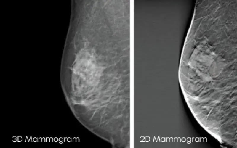
Tomosynthesis represents a cutting-edge advancement in breast imaging technology designed to overcome some limitations of traditional breast imaging. Developed in the early 21st century, tomosynthesis creates a three-dimensional image of the breast by taking X-rays from multiple angles, (sometimes referred to as slices) offering a more comprehensive view of breast tissue layer by layer.
Unlike standard mammography, which captures two-dimensional images, tomosynthesis reduces the impact of overlapping breast tissue, which is a common challenge in interpreting mammogram results. This technique significantly enhances the radiologist's ability to detect breast cancer with higher accuracy, particularly in individuals with dense breast tissue where traditional mammograms might not be as effective.
In addition to improved breast cancer detection rates, tomosynthesis can also decrease the likelihood of false positives and unnecessary callbacks for additional testing. These advantages make it a valuable tool in the early diagnosis and treatment planning of breast cancer.
Which is Better?
Tomosynthesis stands out for its enhanced effectiveness in detecting suspicious lesions. In contrast to the two-dimensional images from standard mammograms, the three-dimensional imaging provided by tomosynthesis delivers more detailed visuals, aiding radiologists in distinguishing between benign and potentially malignant tissue. This screening reduces the occurrence of false positives, especially in patients with dense breast tissue. Although tomosynthesis involves a slightly higher radiation dose than standard mammography, it remains within safe limits, employing the lowest dose necessary for high-quality imaging.
Which One Should You Get?
Who Should Get Mammograms?
- General Screening: Mammograms are recommended for all women over the age of 40 as part of routine breast cancer screening once every two years. The age range can vary based on individual risk factors.
- Those with Average Risk: Women with an average risk of breast cancer, without a personal or strong family history of breast cancer, can opt for traditional mammography as a sufficient screening tool.
- Accessibility and Cost Considerations: Individuals with limited access to the latest technology or who are concerned about the cost or insurance coverage might choose mammography, as it is widely available and often fully covered by insurance.
Who Should Get Tomosynthesis?
- Individuals with Dense Breast Tissue: Tomosynthesis is particularly beneficial for individuals with dense breast tissue. The 3D images provide a clearer view, making it easier to detect cancer.
- Those at Higher Risk of Breast Cancer: Individuals with a higher risk of breast cancer, including those with a family history of the disease, genetic predispositions, or previous breast cancer diagnoses, may benefit from the enhanced imaging that tomosynthesis offers.
- Seeking Detailed Diagnostic Imaging: Anyone seeking the most detailed diagnostic imaging available, especially if previous mammograms have been inconclusive or they need to monitor a known breast condition closely.
Book a Screening Appointment with Canopy Imaging
Tomography and mammography enable individuals to make informed decisions in collaboration with their healthcare providers, ultimately leading to earlier detection and better outcomes in the fight against breast cancer.
Take a vital step towards peace of mind and wellbeing and take charge of your breast health today; book your screening appointment with Canopy Imaging or Auckland Breast Centre, where our expert team provides comprehensive care and state-of-the-art imaging services.