A Three-dimensional (3D) mammogram, also known as breast tomosynthesis, is a type of digital mammography where X-ray machines take pictures of thin slices of the breast to produce a 3D image. This process results in a clearer, more detailed view of the breast tissue, making identifying and characterising abnormalities easier.
Tomosynthesis is similar to how a computed tomography (CT) scanner produces images of structures inside the body.
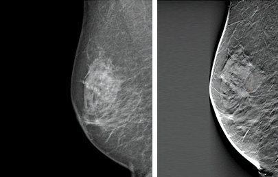
Cancer visible on the right 3D mammogram but not the left 2D mammogram.
In the mammogram image, the glandular and fibrous tissues are white, while the fat is black. However, abnormalities that signal the presence of cancer also appear white and can be mistaken as normal glandular breast tissue, particularly if the breasts are dense.
A 3D tomosynthesis scan takes images, which the computer reconstructs into 1mm slices through the breast. Our specialist Radiologist reads this in conjunction with state-of-the-art 2D digital images.
This scan improves cancer detection and reduces the number of unnecessary biopsies.
Why are we using this technology?
- Increases the cancer detection rate.
- Reduces false negative mammogram results, especially in women with dense breast tissue.
- Reduces the recall rate.
- Reduces unnecessary biopsies.
- Provides a 3D image to the surgeon.
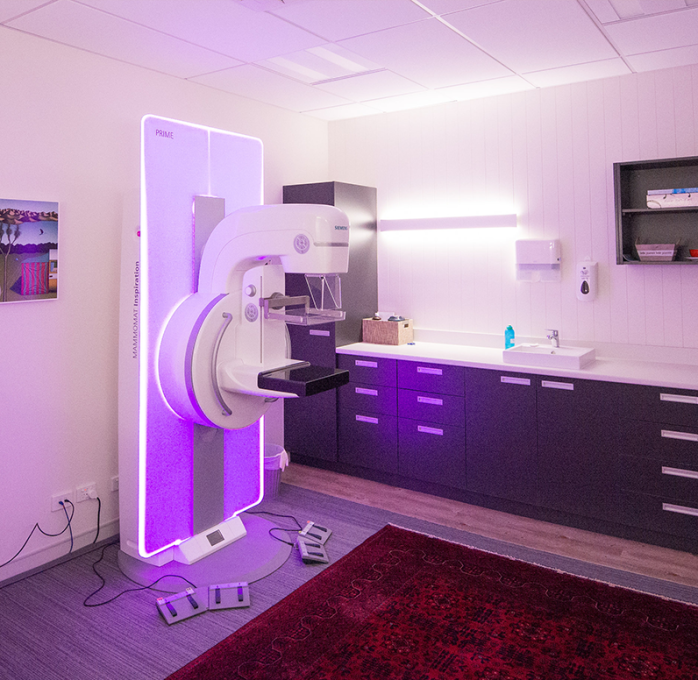
Concerns about 3D Tomosynthesis
What about the radiation dose?
Our dose-reducing software ensures that the total radiation is not significantly different from previous imaging scans.
Is it comfortable?
With new technology, our tomosynthesis system uses less compression and takes about 20 seconds longer to take the image than standard film mammography, designed with patient comfort in mind.
